Wanwarang Teerasamit, Assistant Professor
Abstract
Nowadays, Magnetic resonance imaging (MRI) has an important role for diagnosis of liver lesions due to excellent tissue characterization, radiation-free technique and continuous development of MRI technology, causing an increased use of MRI. This article focuses on basic knowledge of MRI liver interpretation for non-radiologist. The basic techniques including T1-weighted and T2-weighted sequences as well as additional techniques such as 2D dual GRE in-phase and opposed-phase, fat suppression or heavily T2-weighted sequences were introduced. Types of MR contrast agents for liver including extracellular and hepatocyte-specific agents were also described.

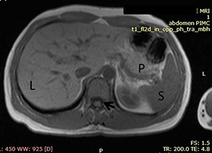
Figure 1 เป็นภาพ MRI ของตับเทคนิค T1W โดยอวัยวะส่วนใหญ่ที่ปรากฏในภาพจะให้ลักษณะ signal intensity ไปในทางhypointense ซึ่งจะเห็นว่าม้าม (spleen=S) ดำกว่าตับ (liver=L) และตับจะดำกว่าตับอ่อน (pancreas=P) โดยลูกศรสีดำชี้ให้เห็น signal intensity ของน้ำไขสันหลังที่เป็นลักษณะ hypointense
DOI https://doi.org/10.30856/th.jhep2019vol2iss1_03
Tags: Miscellaneous


